Full Text:
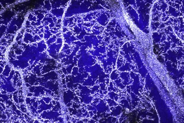
Researchers have discovered a dazzling new method of visualizing neurons that promises to benefit neuroscientists and cell biologists alike: use spectral confocal microscopy to image tissues impregnated with silver or gold. Rather than relying on the amount of light reflecting off metal particles, this novel process involves delivering light energy to silver or gold nanoparticles deposited on neurons and imaging the higher energy levels resulting from their vibrations, known as surface plasmons.
The new process was achieved by using spectral detection on a laser scanning confocal microscope, first made available in the late 1980s and, until now, used most extensively for fluorescent imaging. Paired with such methods, silver- and gold-based cell labeling is poised to unlock new information in a myriad of archived specimens.Image credit: Grant Barthel and Karen Mesce, University of Minnesota and Karen Thompson, Agness Scott College
