Full Text:
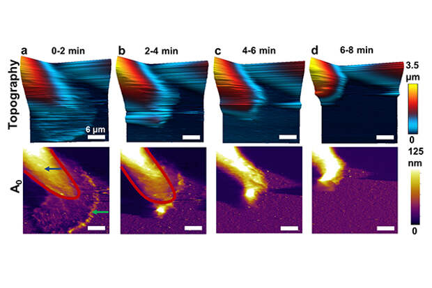
Researchers who developed a high-speed form of atomic force microscopy (AFM) have shown how to image the physical properties of live breast cancer cells for the first time, revealing details about how deactivation of a key protein may lead to metastasis. The new findings also are providing evidence for the mechanisms involved in a cell's response to anti-cancer drugs.
In AFM, a tiny vibrating probe called a cantilever passes over a material, precisely characterizing its topography and physical properties. However, before now the procedure had been too slow to record some quickly changing biological processes in action. Researchers say there is evidence based on this work and previous findings that there might be a mechanical signature to drug resistance.Image credit: Purdue University image/Arvind Raman
